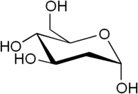
2-Deoxy-D-glucose
 | |
| Names | |
|---|---|
| IUPAC name
(4R,5S,6R)-6-(hydroxymethyl)oxane-2,4,5-triol | |
| Other names
2-Deoxyglucose 2-Deoxy-D-mannose 2-Deoxy-D-arabino-hexose 2-DG | |
| Identifiers | |
| 154-17-6 | |
| 3D model (Jmol) | Interactive image |
| ChemSpider | 388402 |
| ECHA InfoCard | 100.005.295 |
| 4643 | |
| UNII | 9G2MP84A8W |
| |
| |
| Properties | |
| C6H12O5 | |
| Molar mass | 164.16 g/mol |
| Melting point | 142 to 144 °C (288 to 291 °F; 415 to 417 K) |
| Except where otherwise noted, data are given for materials in their standard state (at 25 °C [77 °F], 100 kPa). | |
| | |
| Infobox references | |
2-Deoxy-D-glucose is a glucose molecule which has the 2-hydroxyl group replaced by hydrogen, so that it cannot undergo further glycolysis. As such, it acts to competitively inhibit the production of glucose-6-PO4 from glucose at the phosphoglucoisomerase level.[2] In most cells, glucose hexokinase phosphorylates 2-deoxyglucose, trapping the product 2-deoxyglucose-6-phosphate intracellularly (with exception of liver and kidney); thus, labeled forms of 2-deoxyglucose serve as a good marker for tissue glucose uptake and hexokinase activity. Many cancers have elevated glucose uptake and hexokinase levels. 2-Deoxyglucose labeled with tritium or carbon-14 has been a popular ligand for laboratory research in animal models, where distribution is assessed by tissue-slicing followed by autoradiography, sometimes in tandem with either conventional or electron microscopy.
2-DG is uptaken by the glucose transporters of the cell. Therefore, cells with higher glucose uptake, for example tumor cells, have also a higher uptake of 2-DG. Since 2-DG hampers cell growth, its use as a tumor therapeutic has been suggested, and in fact, 2-DG is in clinical trials [3] A recent clinical trial showed 2-DG can be tolerated at a dose of 63 mg/kg/day, however the observed cardiac side-effects (prolongation of the Q-T interval) at this dose and the fact that a majority of patients' (66%) cancer progressed casts doubt on the feasibility of this reagent for further clinical use.[4] However, it is not completely clear how 2-DG inhibits cell growth. The fact that glycolysis is inhibited by 2-DG, seems not to be sufficient to explain why 2-DG treated cells stop growing [5]
Clinicians have noted that 2-DG is metabolised in the pentose phosphate pathway in red blood cells at least, although the significance of this for other cell types and for cancer treatment in general is unclear.[6]
Work on the ketogenic diet as a treatment for epilepsy have investigated the role of glycolysis in the disease. 2-Deoxyglucose has been proposed by Garriga-Canut et al. as a mimic for the ketogenic diet, and shows great promise as a new anti-epileptic drug.[7] The authors suggest that 2-DG works, in part, by reducing the expression of Brain-derived neurotrophic factor (BDNF), Nerve growth factor (NGF), Arc (protein) (ARC), and Basic fibroblast growth factor (FGF2).[8] Such uses are complicated by the fact that 2-deoxyglucose does have some toxicity.
By combining the sugar 2-Deoxy-D-glucose (2-DG) with fenofibrate, a compound that has been safely used in humans for more than 40 years to lower cholesterol and triglycerides, the entire tumor could effectively be targeted without the use of toxic chemotherapy.[9][10]
2-DG has been used as a targeted optical imaging agent for fluorescent in vivo imaging.[11][12] In clinical medical imaging (PET scanning), fluorodeoxyglucose is used, where one of the 2-hydrogens of 2-deoxy-D-glucose is replaced with the positron-emitting isotope fluorine-18, which emits paired gamma rays, allowing distribution of the tracer to be imaged by external gamma camera(s). This is increasingly done in tandem with a CT function which is part of the same PET/CT machine, to allow better localization of small-volume tissue glucose-uptake differences.
References
- ↑ Merck Index, 11th Edition, 2886.
- ↑ Wick, AN; Drury, DR; Nakada, HI; Wolfe, JB (1957). "Localization of the primary metabolic block produced by 2-deoxyglucose" (PDF). J Biol Chem. 224 (2): 963–969. PMID 13405925.
- ↑ Pelicano, H; Martin, DS; Xu, RH; Huang, P (2006). "Glycolysis inhibition for anticancer treatment". Oncogene. 25 (34): 4633–4646. doi:10.1038/sj.onc.1209597. PMID 16892078.
- ↑ Raez, LE; Papadopoulos, K; Ricart, AD; Chiorean, EG; Dipaola, RS; Stein, MN; Rocha Lima, CM; Schlesselman, JJ; Tolba, K; Langmuir, VK; Kroll, S; Jung, DT; Kurtoglu, M; Rosenblatt, J; Lampidis, TJ (2013). "A phase I dose-escalation trial of 2-deoxy-D-glucose alone or combined with docetaxel in patients with advanced solid tumors". Cancer Chemother. Pharmacol. 71: 523–30. doi:10.1007/s00280-012-2045-1. PMID 23228990.
- ↑ M Ralser, MM Wamelink, EA Struys, C Joppich, S Krobitsch, C Jakobs, H Lehrach Proc Natl Acad Sci U S A, 2008, doi:10.1073/pnas.0803090105
- ↑ JD O'Dea
- ↑ Mireia Garriga-Canut, Barry Schoenike, Romena Qazi, Karen Bergendahl, Timothy J Daley, Rebecca M Pfender, John F Morrison, Jeffrey Ockuly, Carl Stafstrom, Thomas Sutula & Avtar Roopra, "2-Deoxy-D-glucose reduces epilepsy progression by NRSF-CtBP–dependent metabolic regulation of chromatin structure", Nat Neurosci, 9, 1382 - 1387 (2006). doi:10.1038/nn1791 Garriga-Canut, M.; Schoenike, B.; Qazi, R.; Bergendahl, K.; Daley, T. J.; Pfender, R. M.; Morrison, J. F.; Ockuly, J.; Stafstrom, C.; Sutula, T.; Roopra, A. (2006). "2-Deoxy-D-glucose reduces epilepsy progression by NRSF-CtBP–dependent metabolic regulation of chromatin structure". Nature Neuroscience. 9 (11): 1382–1387. doi:10.1038/nn1791. PMID 17041593.
- ↑ Jia Yao, Shuhua Chen, Zisu Mao, Enrique Cadenas, Roberta Diaz Brinton "2-Deoxy-D-Glucose Treatment Induces Ketogenesis, Sustains Mitochondrial Function, and Reduces Pathology in Female Mouse Model of Alzheimer's Disease", PLOS ONE
- ↑ Researchers develop novel, non-toxic approach to treating variety of cancers. ScienceDaily
- ↑ Huaping Liu, Metin Kurtoglu, Clara Lucia León-Annicchiarico, Cristina Munoz-Pinedo, Julio Barredo, Guy Leclerc, Jaime Merchan, Xiongfei Liu, Theodore J. Lampidis (2016). Combining 2-Deoxy-D-glucose with fenofibrate leads to tumor cell death mediated by simultaneous induction of energy and ER stress. Oncotarget, doi:10.18632/oncotarget.9263
- ↑ Kovar, J., Volcheck, W., Sevick-Muraca, E., Simpson, M.A., and Olive, D.M., Analytical Biochemistry, Vol. 384 (2009) 254-262 Download PDF Archived July 13, 2011, at the Wayback Machine.
- ↑ Cheng, Z., Levi, J., Xiong, Z., Gheysens, O., Keren, S., Chen, X., and Gambhir, S., Bioconjugate Chemistry, 17(3), (2006), 662-669