Fimbria of hippocampus
| Fimbria of hippocampus | |
|---|---|
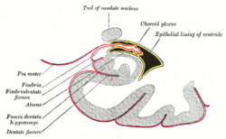
 Coronal section of inferior horn of lateral ventricle. (Fimbria labeled at center left.) | |
| Details | |
| Identifiers | |
| Latin | fimbria hippocampi |
| NeuroNames | hier-169 |
| NeuroLex ID | Fimbria of hippocampus |
| TA |
A14.1.09.238 A14.1.09.332 |
| FMA | 83728 |
With regard to the brain, the fimbria is a prominent band of white matter along the medial edge of the hippocampus.
Structure
The fimbria is an accumulation of myelinated axons (mostly efferent) that first collect on the ventricular surface of the hippocampus as the alveus (a thin layer resembling an inverted trough).
Relations
Near the splenium the fimbria separates from the hippocampus as the crus fornicis.
Additional images
.jpg) Diagram of hippocampus
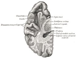
Diagram of hippocampus Section of brain showing upper surface of temporal lobe.
 Scheme of rhinencephalon.
 Posterior and inferior cornua of left lateral ventricle exposed from the side.
 Inferior and posterior cornua, viewed from above.
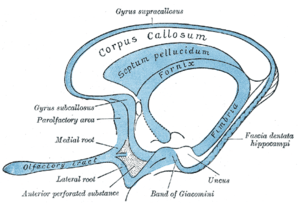
 Diagram of the fornix.
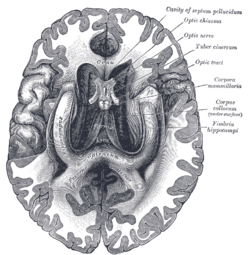
 The fornix and corpus callosum from below.
| Wikimedia Commons has media related to Fimbria of hippocampus. |
This article is issued from Wikipedia - version of the 12/9/2014. The text is available under the Creative Commons Attribution/Share Alike but additional terms may apply for the media files.