Lateral spinothalamic tract
| Lateral spinothalamic tract | |
|---|---|
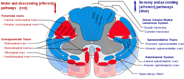
 Lateral spinothalamic tract is labeled in blue at lower right. | |
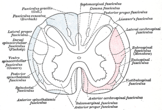
 Diagram of the principal fasciculi of the spinal cord. | |
| Details | |
| Identifiers | |
| Latin | tractus spinothalamicus lateralis |
| TA | A14.1.02.225 |
| FMA | 73965 |
The lateral spinothalamic tract (or lateral spinothalamic fasciculus), which is a part of the anterolateral system, is a bundle of sensory axons ascending through the white matter of the spinal cord, carrying sensory information to the brain. It carries pain,fine touch and temperature sensory information (protopathic sensation) to the thalamus. It is composed primarily of fast-conducting, sparsely myelinated A delta fibers and slow-conducting, unmyelinated C fibers. These are secondary sensory neurons which have already synapsed with the primary sensory neurons of the peripheral nervous system in the posterior horn of the spinal cord (one of the three grey columns).
Together with the anterior spinothalamic tract, the lateral spinothalamic tract is sometimes termed the secondary sensory fasciculus or spinal lemniscus.
Anatomy
The neurons of the lateral spinothalamic tract originate in the spinal ganglia. They project peripheral processes to the tissues in the form of free nerve endings which are sensitive to molecules indicative of cell damage. The central processes enter the spinal cord in an area at the back of the posterior horn known as the posterolateral tract. Here, the processes ascend approximately two levels before synapsing on second-order neurons. These secondary neurons are situated in the posterior horn, specifically in the Rexed laminae regions I, IV, V and VI. Region II is primarily composed of Golgi II interneurons, which are primarily for the modulation of pain, and largely project to secondary neurons in regions I and V. Secondary neurons from regions I and V decussate across the anterior white commissure and ascend in the (now contralateral) lateral spinothalamic tract. These fibers will ascend through the brainstem, including the medulla oblongata, pons and midbrain, as the spinal lemniscus until synapsing in the ventroposteriorlateral (VPL) nucleus of the thalamus. The third order neurons in the thalamus will then project through the internal capsule and corona radiata to various regions of the cortex, primarily the main somatosensory cortex, Brodmann areas 3, 1, and 2.
Function
The fibers of the lateral spinothalamic tract conduct information about temperature and pain.
See also
References
This article incorporates text in the public domain from the 20th edition of Gray's Anatomy (1918)